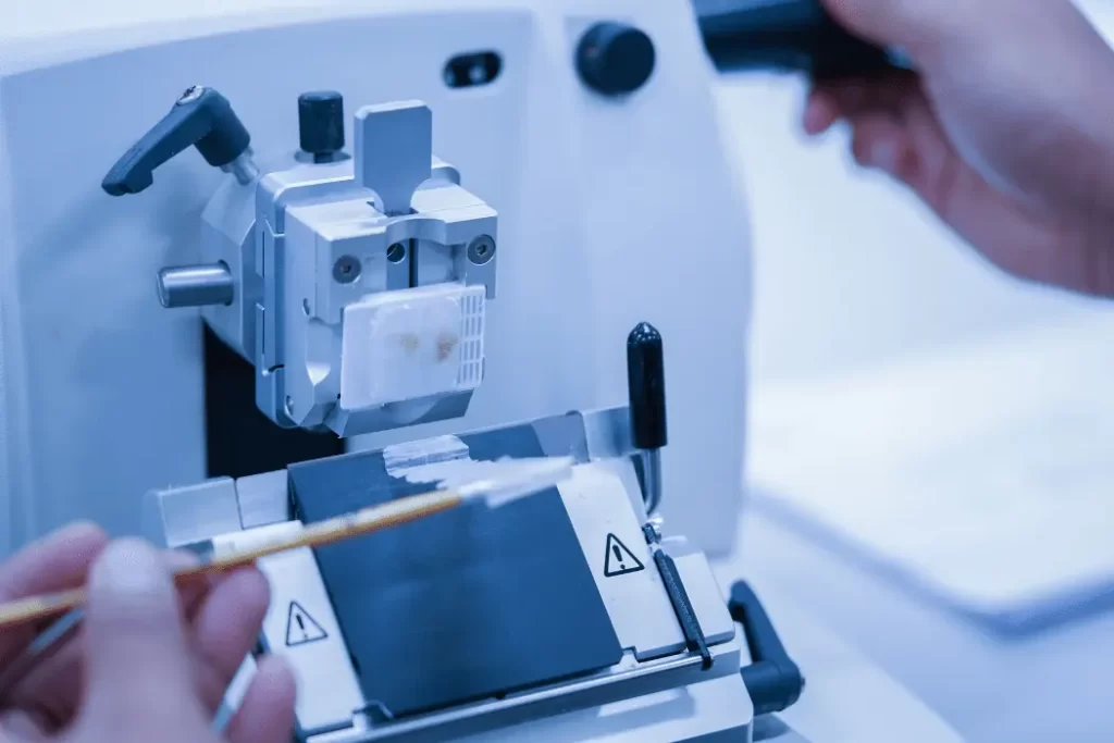
Imagine a world where medical professionals could effortlessly obtain thin and precise tissue slices for accurate diagnosis and research purposes. Thanks to the incredible tool known as a microtome, this vision has become a reality. In this article, we will explore the art of microtome mastery, discussing its importance, techniques, and benefits. So, let’s delve into the fascinating world of perfecting precise tissue slicing.
Microtomy, the art of slicing extremely thin sections of tissues, has revolutionized the field of histology and pathology. By enabling researchers and medical professionals to examine tissue samples under a microscope, microtomes have become indispensable tools in various scientific disciplines. Microtome mastery is the ultimate source of knowledge for perfecting precise tissue slicing, unlocking a wealth of insights in the field of histology and pathology. In this article, we will delve into the mastery of microtomes, focusing on the techniques and considerations necessary for obtaining precise tissue slices.
The Significance of Microtome Mastery
Mastering the art of microtome operation is crucial for ensuring accurate results in histological research and medical diagnoses. The quality of tissue slices greatly impacts the interpretation of microscopic images and subsequent analysis. By achieving optimal sectioning, medical professionals can unlock valuable insights into cellular structures, identify abnormalities, and advance medical knowledge.
Understanding the Microtome
A microtome is a precision instrument used to slice thin sections from biological specimens. It allows for controlled cutting of tissues with thicknesses ranging from a few micrometers to several millimeters. Modern microtomes feature advanced mechanisms for precise control of section thickness and sample orientation.

Types of Microtomes
There are several types of microtomes available, each suited for specific applications. Let’s explore the most common ones:
Rotary Microtome
The rotary microtome is the traditional workhorse in histology laboratories. It employs a rotary mechanism to cut tissue samples mounted on a specimen holder. The section thickness is determined by adjusting the position of the blade and the advancement mechanism.
Vibrating Microtome
Vibrating microtomes use a vibrating blade to slice through the tissue, allowing for smooth and precise sectioning. This type of microtome is particularly useful for delicate samples and specialized applications.
Cryostat Microtome
Cryostat microtomes are specifically designed to section frozen tissue samples. They maintain low temperatures, enabling the slicing of tissues without the need for fixation or embedding. Cryostat microtomes are invaluable in studies requiring rapid sectioning of fresh-frozen specimens.
Choosing the Right Microtome
Selecting the appropriate microtome depends on the specific research or diagnostic requirements. Factors to consider include the type of samples, desired section thickness, level of automation, and budget constraints. Consulting with experienced professionals and considering user reviews can help make an informed decision.
Preparation and Safety Measures
Before starting the microtome slicing process, it is essential to prepare the tissue samples adequately. This involves proper fixation, embedding in a supporting medium, and trimming the sample blocks to the desired size. Additionally, wearing personal protective equipment, such as gloves and safety goggles, ensures operator safety during the sectioning procedure.
The Art of Sectioning
Achieving precise tissue sections requires a systematic approach. Here are the key steps involved in the sectioning process:
Fixation and Embedding
Fixation preserves the tissue structure and prevents degradation. After fixation, the sample is embedded in a solid medium, such as paraffin wax or resin, providing stability during sectioning.
Block Trimming
Before sectioning, the embedded tissue block needs to be properly trimmed to obtain a flat surface for slicing. Trimming ensures consistent and parallel sections.
Sectioning Technique
Using the microtome, the tissue block is sliced into thin sections. The operator adjusts the thickness, advances the sample, and carefully collects the sections on glass slides or other suitable surfaces.
Collecting and Mounting Slices
The obtained tissue slices are delicately transferred onto glass slides and allowed to dry. Once dry, they can be stained using various techniques to enhance visualization under a microscope.
Enhancing Slice Quality
To obtain high-quality tissue slices, certain considerations come into play:
Temperature Control
Maintaining the appropriate temperature during sectioning is crucial. Different types of tissues require specific temperature ranges to achieve optimal sectioning results.
Blade Selection and Maintenance
Choosing the right blade type, sharpness, and properly maintaining it are essential for achieving clean and accurate cuts. Dull or damaged blades can negatively impact slice quality.
Staining and Visualization
Staining tissue slices with dyes enhances contrast and facilitates the identification of cellular components. Various staining techniques exist, enabling different structures to be highlighted.
Applications of Precise Tissue Slicing
The applications of precise tissue slicing are vast and span multiple scientific disciplines. Some notable applications include:
- Histopathology: Analysis of tissue samples for diagnosing diseases.
- Research: Investigating cellular structures and functions.
- Drug Discovery: Evaluating the effects of potential therapeutic compounds on tissues.
- Forensics: Examining postmortem tissues for evidence collection.
Troubleshooting Common Issues
During the sectioning process, several common issues may arise. Let’s explore a few and their potential solutions:
Wrinkles and Folds
Wrinkles and folds on tissue sections can result from poor sample preparation or incorrect handling. Ensuring proper embedding and gentle handling of the samples can help minimize these issues.
Chatter Marks
Chatter marks appear as uneven and irregular lines on the tissue sections. They can be caused by vibration or instability in the microtome setup. Stabilizing the microtome and optimizing cutting conditions can alleviate this problem.
Knife Lines
Knife lines manifest as visible lines or artifacts on the tissue sections. They can occur due to improper blade alignment or contamination. Regular blade alignment checks and maintaining a clean cutting environment can mitigate knife line formation.
The Future of Microtome Technology
As technology advances, microtomes continue to evolve. Future developments may include increased automation, integration with digital imaging systems, and enhanced precision through advanced blade control mechanisms. These advancements will further streamline the process of precise tissue slicing, making it even more accessible and accurate.
Conclusion
Mastering the art of microtome operation is essential for obtaining precise tissue slices. Through a systematic approach and attention to detail, medical professionals and researchers can unlock valuable insights into cellular structures and advance their respective fields. Whether it’s for diagnostic purposes or scientific investigations, microtome mastery plays a vital role in the pursuit of knowledge and innovation.
FAQs on Microtome
Q: What is a microtome?
Answer: A microtome is a precision instrument used to slice thin sections of tissues for microscopic examination and analysis.
Q: What is the purpose of using a microtome?
Answer: The primary purpose of using a microtome is to obtain thin and uniform tissue sections for various applications in histology, pathology, and research.
Q: What are the different types of microtomes?
Answer: There are several types of microtomes, including rotary microtomes, vibrating microtomes, and cryostat microtomes, each with its own specific functions and advantages.
Q: How do microtomes work?
Answer: Microtomes work by holding a tissue sample and precisely cutting it into thin slices using a blade or vibrating mechanism, allowing for detailed examination under a microscope.
Q: Are there any safety considerations when using a microtome?
Answer: Yes, safety precautions, such as wearing personal protective equipment (PPE) like gloves and safety goggles, should be followed to ensure operator safety during the microtome operation.


